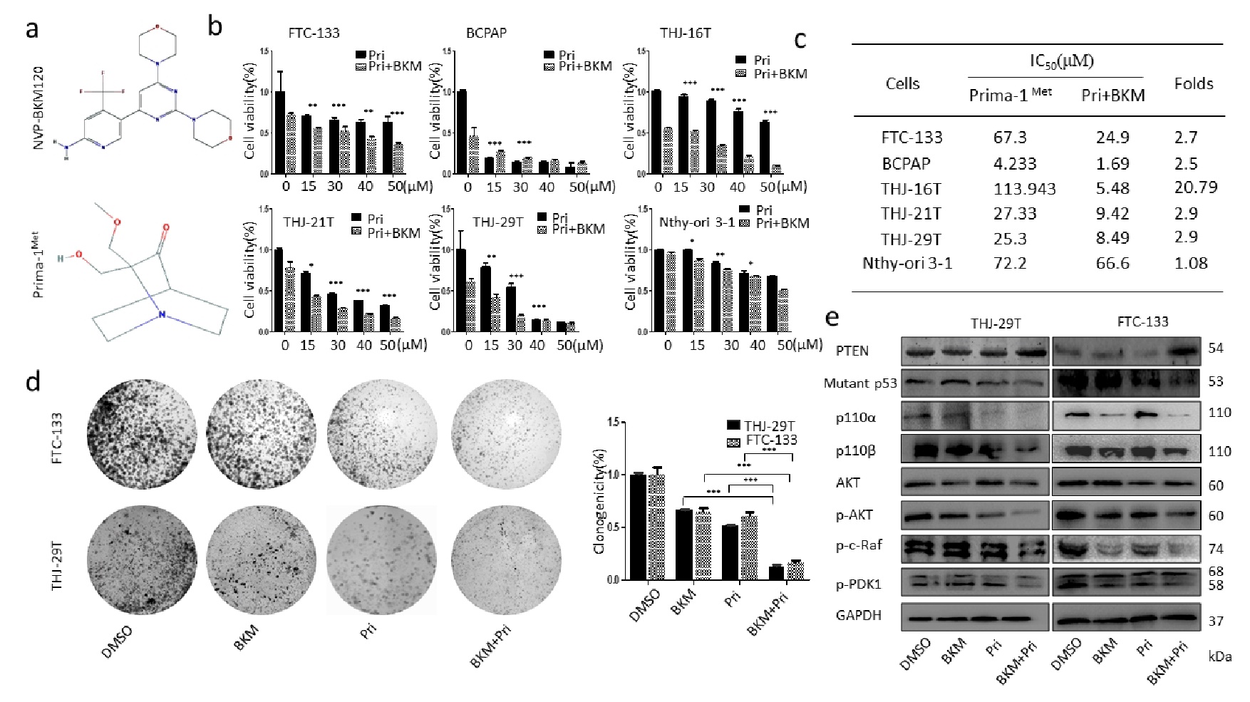
Fig. 1. Effect of the combination treatment of NVP-BKM120 and Prima-1Met on cell proliferation in thyroid cancer cells. (a) Chemical structure of NVP-BKM120 and Prima-1Met. (b) Thyroid cancer cells (THJ-16T, THJ- 21T, THJ-29T, FTC-133, BCPAP) and normal thyroid cells (Nthy-ori-3-1) were treated with the indicated doses of Prima-1Met alone or in combination with BKM120 (1 µM) for 48 hours, and cell viability was determined by MTT assay. (c) The IC50 values of Prima-1Met were determined for cell viability inhibition in cells treated with Prima-1Met alone or in combination with BKM120 (1 µM). The data were presented as mean ± SD of three independent experiments. *P<0.05 and **P<0.01, significant differences compared to the control groups. (d) Colony formation was shown for two thyroid cancer cell lines (THJ-29T, FTC-133) treated with BKM120 (1 µM), Prima-1Met (30 µM) or the two in combination for 48 hours. (e) THJ-29T and FTC-133 cells were treated with BKM120 (1 µM), Prima-1Met (30 µM) or the two in combination for 48 hours. The protein levels of p110a, p110ß, total and phosphorylated Akt, p-PDK1, p-c-Raf, p-PTEN, and mutant p53 were analyzed by Western blot. ß-actin served as the loading control. All the experiments were repeated 3 times, and the data were shown as mean ± SD. P-values were calculated by student’s t-test.